FIXING CELLS WITH PRIMARY FIXATIVE:
(1) Take cells (12 well plates) from tissue culture incubator and chill them at 4 degrees celsius for at least 3 hours.
You can perform the fixation procedure in a cold room. Alternatively, if you want to do it on your bench, make sure you move
fast and keep all solutions cold.
(2) Prepare fresh primary fixative, 4% Glutaraldehyde in 150 mM HEPES pH7.5 (Remember to do it under the hood). Filter
the solution through a 0.2 micron filter and chill on ice. Alternatively, prepare the day before and store air-tight at 4
degrees celsius.
(3) Rinse cells by transfering Transwells sequentially through 5 beakers of chilled PBS (4 degrees celsius).
(4) Place the Transwell in a clean 12 well plate on ice. Qucikly but gently add 3 mL of cold primary fixative (4% Glutaraldehyde
in 150 mM HEPES pH7.5) to the apical well of Transwell. Let the fixative overflow into the basal well. You may want to do
this under the hood.
(5) Repeat steps (3) and (4) for each Transwell.
(6) Place Transwells/plate in an air-tight container. Let fix overnight at 4 degrees celsius.
QUENCHING THE PRIMARY FIXATIVE:
(1) Prepare quenching solution, 50 mM glycine in 100 mM HEPES pH7.5. Keep at 4 degrees celsius.
(2) Remove primary fixative from Transwells.
(3) Quench cells by transfering Transwells sequentially through 3 beakers of cold quenching solution. Let cells quench
in 12 well plate for an additional 15 min on ice.
(4) Rinse cells by sequentially trasfering Transwells to 5 beakers of cold distilled water. Keep in water on ice.
FIXING CELLS WITH SECONDARY FIXATIVE:
(1) Prepare fresh secondary fixative, 1% Osmium tetroxide in 2.5% potassium ferrocyanide. Use immediatedly. Osmium
is very volatile and toxic. Must work under hood.
(2) Remove liquid from Transwell, added 0.5 mL secondary fixative to the apical well, 1 mL to the basal well. Let fix
on ice for 2 hours in Dark (cover with foil).
(3) Remove apical and basal secondary fixative with pipet tip. Transfer osmium solution to Toxic Waste Container.
(4) Rinse cells by transfering Transwells sequentially through 5 beakers of cold distilled water. Keep in water on ice.
EN BLOC STAINING WITH URANYL ACETATE:
(1) Prepare 2% uranyl acetate in distilled water. Filter through 0.2 µm pore membrane.
(2) Remove liquid from Transwell, added 0.5 mL 2% uranyl acetate to the apical well, 1 mL to the basal well. Let stain
at room temperature for 2 hours.
(3) Remove apical and basal staining solution with pipet tip. Transfer uranyl solution to proper Waste Container.
(4) Rinse cells by sequentially trasfering Transwells to 5 beakers of cold distilled water. Keep in water on ice.
DEHYDRATION AND EMBEDDING:
(1) At room temperature. Dehydrate cells by transfering Transwells sequentially through beakers of 50%, 75%, 95%, 100%,
100%, 100% ethanol. Let sit in 100 % ethanol in 12 well plate for 10 min.
(2) Very quickly remove as much ethanol from the apical well as possible. Immediately add embedding medium/Araldite
Epoxy. DO NOT LET CELL DRY.
(3) Bake at 65 degrees celsius for at least 48 hours.
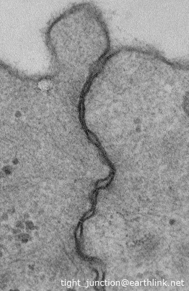
SECTIONING THE CELLS:
(1) Cut the cells from apical to basal to minimize curling of the sections.
(2) Cut 50-70 nm sections for examination of junctional structures. Post-section stain with lead citrate on carbon coated
EM grid.
(3) Cut 150 nm for thick-section light microscopy. Post-section stain with 1% methylene blue on glass slide.